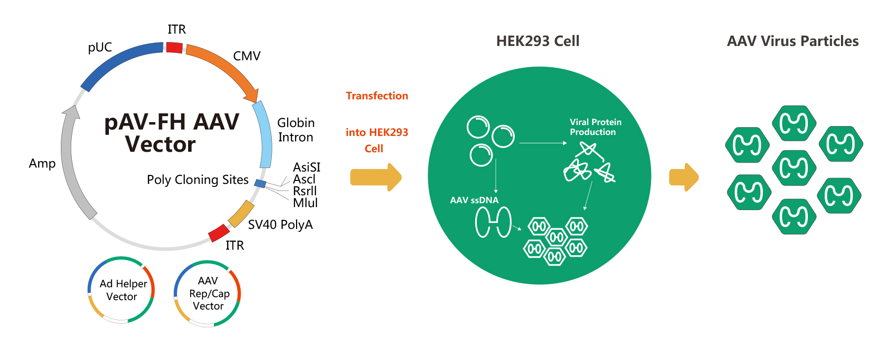
WZ Biosciences adopts modified AAV Helper-Free System for AAV packaging. The schematic diagram of AAV packaging system is below:
AAV Packaging Procedure
WZ Biosciences provides a full suite of services ranging from designing, cloning, packaging to expression analysis. Also, WZ Biosciences offers the complete AAV vectors and AAV Expression Systems that can be used to express human/mouse/rat ORFs, lncRNAs, circRNAs, shRNAs, CRISPR/gRNA in vitro and in vivo studies. Working with WZ, you will get high titer, high purity and high stability viral stock. Figure below is the flow chart for AAV production.
AAV Purification
WZ Biosciences purify AAV by Iodixanol Gradient Ultracentrifugation. Iodixanol is a contrast agent, and has been subjected to clinical testing. Iodixanol is non-ionic, non-toxic to cells and metabolically inert. Currently, Iodixanol is widely used for viral purification. Its principle is that particles with different sedimentation rates will lay as different narrow zone under a certain centrifugal force. Figure below is the example of iodixanol gradients before and after centrifugation.
AAV Titering
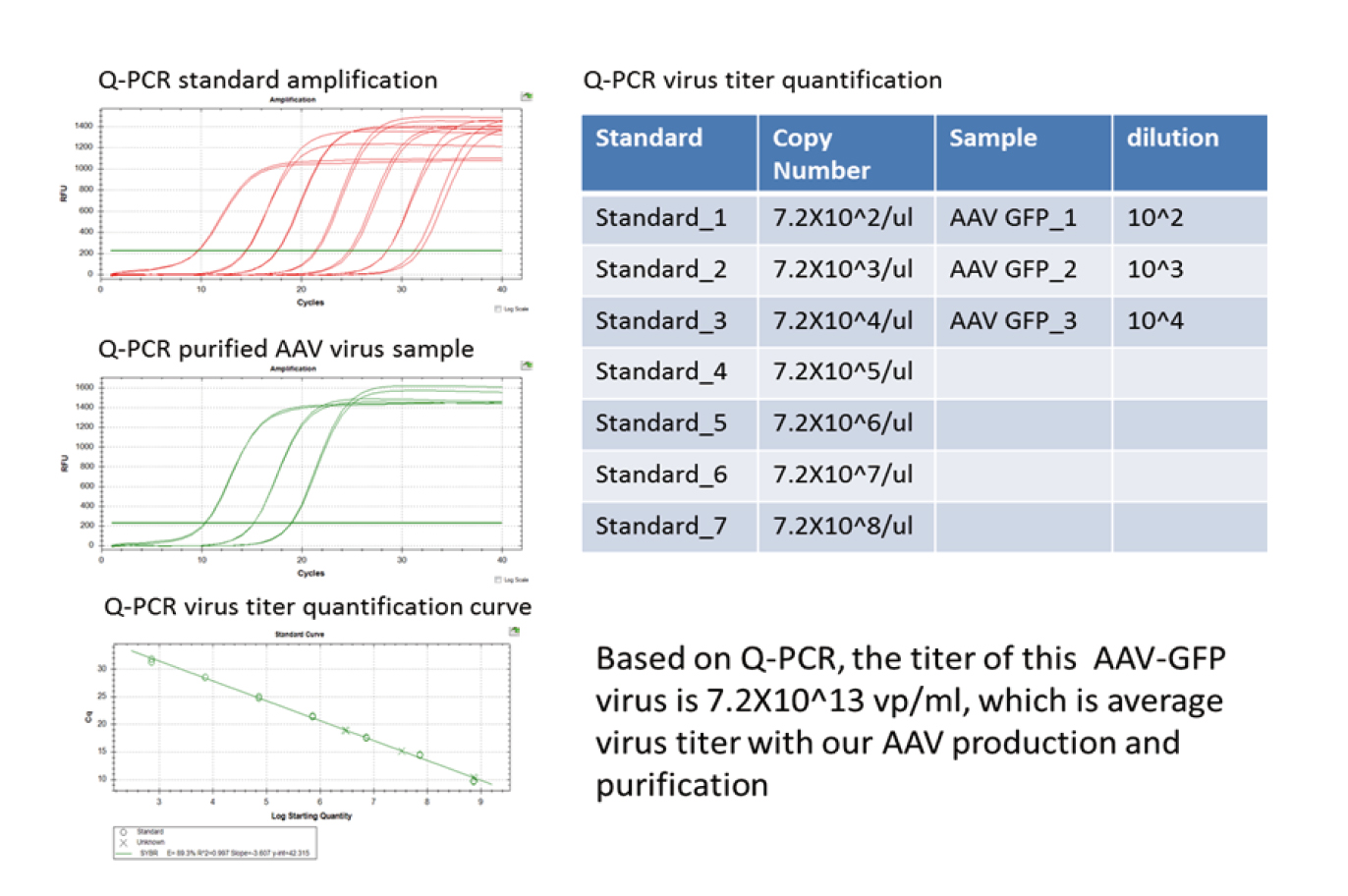
WZ Biosciences uses a quantitative polymerase chain reaction (qPCR) for measuring physical titer(viral particles) for all AAVs. We performe qPCR assay with specific primers targeting AAV genome. The viral titer is quantified by comparison to a standard curve of a plasmid sample of known concentration. Our AAV-GFP tittering methodology is shown below.
AAV Purity Testing
Our AAV purity is assessed by SDS-PAGE. AAV capsid proteins are composed of three structural proteins (VP1, VP2 and VP3) which assemble at a ratio of approximately 1:1:10. Therefore, a high purity AAV sample should essentially show only VP1, VP2, and VP3, and there are no visible protein contaminants. Figure below represents the SDS-PAGE analysis of various high purity AAV serotypes, and the predominant bands are VP1 (82kDa)、VP2 (72kDa) and VP3 (62kDa).
AAV Structure
AAV has a diameter of 20-26nm. Its diameter is 1/5 of adenovirus and 1/4 of lentivirus. It has a regular icosahedron structure and can infect both dividing and non-dividing cells.
AAV Genome
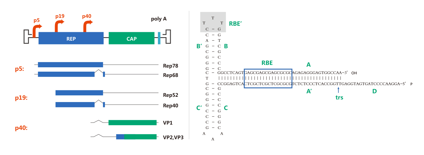
AAV has a linear single-stranded DNA genome of about 4.7 kb. Its genome contains inverted terminal repeats (ITRs) at both ends of the DNA strand and two open reading frames (ORFs): rep and cap. These ITRs are important for AAV genome replication and packaging; The Rep gene encodes regulatory proteins, which are required for viral genome replication and integration, while Cap gene encodes structural proteins VP1, VP2 and VP3 in a ratio of approximately 1:1:10 (VP1:VP2:VP3). Based on the structure of AAV genome, recombinant AAV is designed by replacing all AAV protein-coding sequences between the ITRs with exogenous gene expression cassettes. The only sequences of viral origin are two ITRs, which are necessary to guide genome replication and packaging during AAV preparation. The complete removal of viral coding sequences maximizes the packaging capacity of AAV and contributes to their low immunogenicity and cytotoxicity when delivered in vivo.
AAV Serotypes
Currently, 12 distinct human AAV serotypes (AAV1 to AAV12) and more than 100 non-human primate AAV serotypes have been identified. Different AAV serotypes have different capsid protein spatial structure, sequence and tissue specificity, therefore the cell surface receptors that AAVs recognize and bind are significant different. That’s the reason why AAV serotypes have varying cell type, tissue tropisms and transduction efficiencies. Table 1: Known cellular receptors for nine human AAV serotypes. Table 2:A summary of the tissue tropism of AAV serotypes.
|
Serotype
|
Glycan recognitiona
|
Coreceptor
|
|
AAV1
|
Neu5Aca2-3GalNAcβ1-4GlcNAc
|
Unknown
|
|
AAV2
|
6-O-and N-sulfated heparin
|
Fibroblast / hepatocyte growth factor receptor;
laminin receptor; integrin αVβ5 and α5β1
|
|
AAV3
|
2-O-and N-sulfated heparin
|
Hepatocyte growth factor receptor; Laminin receptor
|
|
AAV4
|
Galβ1-4GlcNAcβ1-2Manα1-6Manβ1-4GlcNAcβ1-4GlcNAc
|
Unknown
|
|
AAV5
|
Neu5Acα2-3(6S)Galβ1-4GlcNAc
|
Platelet-derived growth factor receptor
|
|
AAV6
|
Neu5Acα2-3GalNAcβ1-4GlcNAc; N-sulfated heparin
|
Epidermal growth factor receptor
|
|
AAV7
|
Unknown
|
Unknown
|
|
AAV8
|
Unknown
|
Laminin receptor
|
|
AAV9
|
Galactose
|
Laminin receptor
|
|
Tissue
|
Optimal Serotype
|
|
CNS
|
AAV1,AAV2,AAV4,AAV5,AAV8,AAV9
|
|
Heart
|
AAV1,AAV8,AAV9
|
|
Kidney
|
AAV2
|
|
Liver
|
AAV7,AAV8,AAV9
|
|
Lung
|
AAV4,AAV5,AAV6,AAV9
|
|
Pancreas
|
AAV8
|
|
Photoreceptor Cells
|
AAV2,AAV5,AAV8
|
|
RPE(Retinal Pigment Epithelium)
|
AAV1,AAV2,AAV4,AAV5,AAV8
|
|
Skeletal Muscle
|
AAV1,AAV6,AAV7,AAV8,AAV9
|
AAV Transduction Pathway
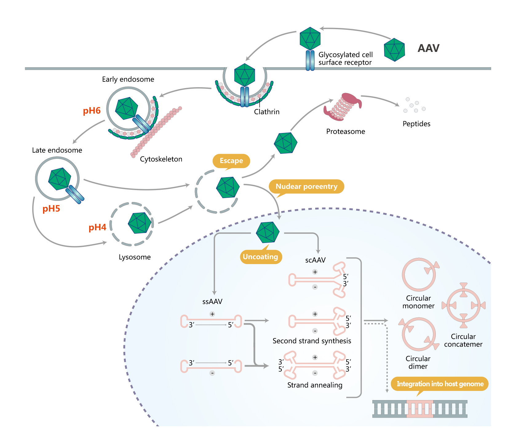
The process of AAV infection begins with recogning and binding to the cell surface glycosylated receptors. This triggers internalization of AAV via clathrin-mediated endocytosis. AAV then traffics through the cytosol mediated by the cytoskeletal network. Owing to the somewhat low pH environment of the endosome, the VP1/VP2 region undergoes a conformational change. Following endosomal escape, AAV undergoes transport into the nucleus and uncoating. AAV can also undergo proteolysis by the proteasome. After the single- to double-stranded DNA conversion, forming circularized episomal genomes that can persist in the nucleus.
ssAAV & scAAV
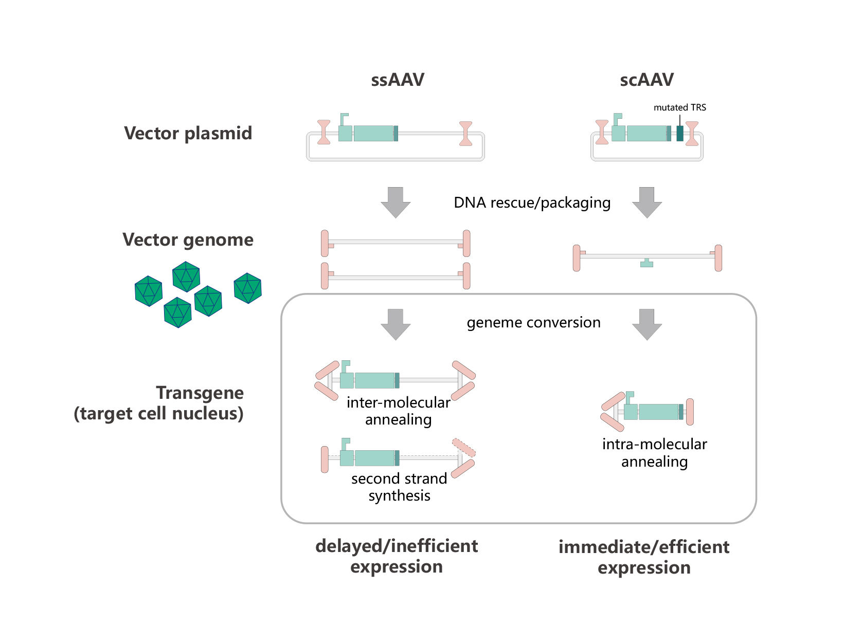
Currently, there are two classes of AAV in use: single-stranded AAV (ssAAV) and self-complementary AAV (scAAV). scAAV is developed based on two principles: one is theorical foundation that virus particles, whether scAAV or rAAV, can be encapsulated with diploid or even tetraploid AAV genomic DNA; the other is structural foundation that wtAAV ITR forms a characteristic T-shaped hairpin structure(see AAV genome section). After entering the nucleus and decoating, ssAAV genomes with sense (plus) and anti-sense (minus) orientations are packaged equally well. ssDNA must be converted by host polymerase or intermolecular annealing into a double-stranded form before transcription of the transgene can take place. Because scAAVs are already double-stranded by design, they can immediately undergo transcription. Figure below is the process of ssAAV and scAAV delivering foreign genes to target cells.
|
1. Complete Vectors:
|
Over 30 tissue specific promoters and different reporters (GFP, RFP or luciferase and so on) available for AAV cloning.
|
|
|
|
2. Quick Delivery:
|
All AAV cDNA & mirRNA cloning vectors are compatible with our Entry & MIR clones MCS– choose from 18,000 ORF clones & 1300 microRNA clones.
|
|
|
|
3. High Titer:
|
titer=10E12-13 vp/ml, up to 10E14 vp/ml.
|
|
|
|
4. Excellent Service:
|
Based on your research goal, our professional technical staffs will design experimental program and manufacture the most ideal AAV particles for you.
|

|
《Adeno-associated virus technical manual》
|
|
|
1. What is adeno-associated virus (AAV) & recombinant AAV(rAAV) ?
The adeno-associated virus (AAV) is a small, icosahedral and non-enveloped virus that belongs to Parvoviridae family. Helper virus such as Adenovirus or Herpes virus is usually required for a productive infection to occur. AAV does not encode its own polymerase so its replication process relies on host cell polymerase activities.
Recombinant AAV is the artificial AAV without any AAV rep and cap genes which encode viral replication and structural proteins, respectively. In rAAV, rep and cap are replaced with a gene or construct of interest flanked by the ITRs for replication and packaging. Efficient packaging of rAAV can be performed with constructs ranging from 4.1 kb to 4.9 kb in size.
2. What’s the Biosafety requirement for using AAV?
Recombinant AAV constructs produced in the absence of a helper virus and encodes no tumorigenic gene can be handled in Biosafety Level 1(BSL-1) facility. Otherwise, it should be handled as biohazardous material under Biosafety Level 2 (BSL-2) containment.
3. Is the recombinant AAV safe?
To date, AAV is not linked to any human disease. For wild type AAV, replication is at extremely low efficiency, without the presence of helper virus, such as adenovirus. The recombinant AAV (rAAV) composed by several plasmids (cis plasmid, Helper plasmid, rep/Cap plasmid). Cis plasmid and Helper do not share any regions of homology with the rep/cap-gene containing plasmid, the likelihood for a recombinant AAV to replicate is theoretically impossible.
4. What’s the cloning capacity for recombinant AAVs?
AAV has a packaging capacity of ~4.7Kb. When the length of inserted DNA between the 2 ITRs is close the maximal allowed, i.e., 4-4.4Kb, the packaging efficiency decreases significantly. For instance, for gene over-expression from cDNA, since the CMV-poly(A) element is about 1Kb, so the maximal allowable cDNA length is about 3K, whereas if GFP co-expression ( about 2Kb) is considered, the allowable capacity is about 1-1.2 Kb.
5. What AAV serotype do you provide?
We currently provide AAV serotype AAV1,AAV2,AAV5,AAV6,AAV7,AAV8 &,AAV9,AAV rh10,AAV retro,AAV ANC80,AAV DJ & AAV DJ-8,AAV PhpB & AAV PhpeB,AAV 7m8 and AAV shh10.
6. What are the advantages of gene delivery by rAAV?
rAAV has the capacity to produce high titer virus in dividing and non-dividing cells and potential for long-term gene transfer with minimum immnunogenicity.
7. How stable are AAV vectors? How should they be stored?
Purified AAV vectors are highly stable at temperatures of 4 C or less. We recommend aliquoting upon receipt and storing stock at -80C for long term storage.
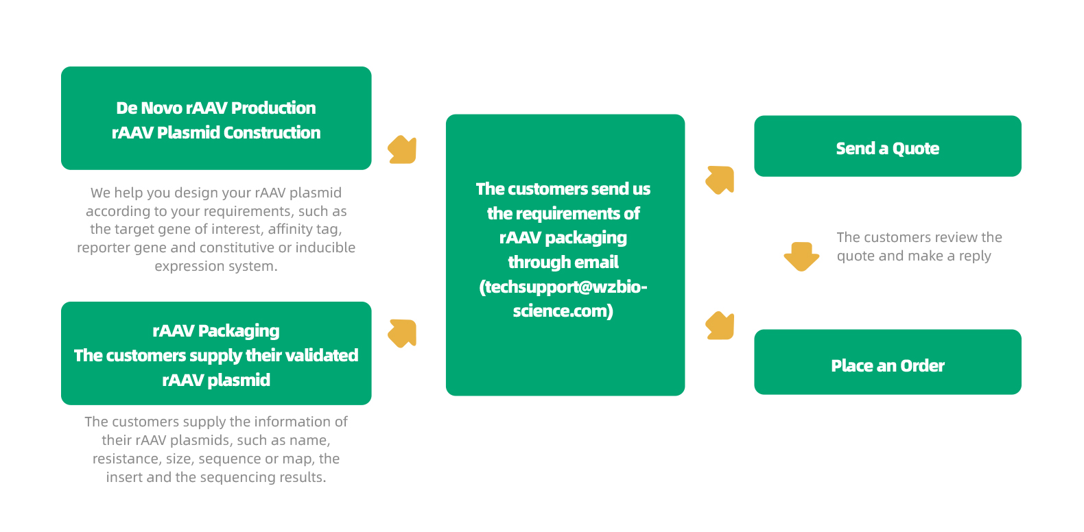
8. What does a customer need to provide for the custom AAV service? & Expected yield?
Please provide us with plasmid DNA for the specified gene, vector map and its sequence information.
For gene silencing service, we need the exact shRNA sequence to be constructed into recombinant AAV vector if no plasmid available.
Normally, expected yield is between 1x10^12-13 vp/ml, with about 500 ul viral stock will be given. However, special quantity/titer can be made upon request.
Central Nervous System(CNS):
|
1.
|
Nature Communications. (IF=12.121). Xu, et.al. (2020). CircGRIA1 shows an age-related increase in male macaque brain and regulates synaptic plasticity and synaptogenesis.
|
|
|
2.
|
Science Advances. (IF=13.116). Sun, et.al. (2020). Development of a CRISPR-SaCas9 system for projection- and function-specific gene editing in the rat brain.
|
|
|
3.
|
The Journal of Neuroscience. (IF=5.673). Zingg B, et.al. (2020). Synaptic Specificity and Application of Anterograde Transsynaptic AAV for Probing Neural Circuitry.
|
|
|
4.
|
Molecular Psychiatry. (IF=12.384). li, et.al. (2020). Programmed cell death 4 as an endogenous suppressor of BDNF translation is involved in stress-induced depression.
|
|
|
5.
|
Biological Psychiatry. (IF=12.095). Zhang, et.al. (2020). Reduced neuronal cAMP in the nucleus accumbens damages blood-brain barrier integrity and promotes stress vulnerability.
|
|
|
6.
|
Cell Reports. (IF=8.109). Ma, et.al. (2019). Spontaneous Pain Disrupts Ventral Hippocampal CA1-Infralimbic Cortex Connectivity and Modulates Pain Progression in Rats with Peripheral Inflammation.
|
|
|
7.
|
Neuron. (IF=14.415). Feng, et.al. (2019). A Genetically Encoded Fluorescent Sensor for Rapid and Specific In Vivo Detection of Norepinephrine.
|
|
|
8.
|
Cell. (IF=38.637). Sun, et.al. (2018). A Genetically Encoded Fluorescent Sensor Enables Rapid and Specific Detection of Dopamine in Flies,Fish, and Mice.
|
|
|
9.
|
Behavioural Neurology. (IF=2.093). Qu, et.al. (2018). MST1 Suppression Reduces Early Brain Injury by Inhibiting the NF-κB/MMP-9 Pathway after Subarachnoid Hemorrhage in Mice.
|
|
|
10.
|
Neuron. (IF=14.415). Zingg B, et.al. (2017). AAV-Mediated Anterograde Transsynaptic Tagging: Mapping Corticocollicular Input-Defined Neural Pathways for Defense Behaviors.
|
|
|
1.
|
Proc. Natl. Acad. Sci. U.S.A. (PNAS). (IF=9.412). Zhang, et.al. (2020). Hepatic neddylation targets and stabilizes electron transfer flavoproteins to facilitate fatty acid β-oxidation.
|
|
|
2.
|
Journal of Hepatology. (IF=20.582). She, et.al. (2019). PSMP/MSMP promotes hepatic fibrosis through CCR2 and represents a novel therapeutic target.
|
|
|
3.
|
Theranostics. (IF=8.579). Liu, et.al. (2019). Suppression of YAP/TAZ-Notch1-NICD axis by bromodomain and extraterminal protein inhibition impairs liver regeneration.
|
|
|
4.
|
Molecular Cell. (IF=15.584). Wan, et.al. (2019). Pacer is a mediator of mTORC1 and GSK3-TIP60 signaling in regulation of autophagosome maturation and lipid metabolism.
|
|
|
5.
|
Experimental Cell Research. (IF=3.383). Li, et.al. (2018). Brg1 promotes liver fibrosis via activation of hepatic stellate cells.
|
|
|
6.
|
Molecular Cell. (IF=15.584). Wan, et.al. (2018). mTORC1-Regulated and HUWE1-Mediated WIPI2 Degradation Controls Autophagy Flux.
|
|
|
7.
|
Molecular Cell. (IF=15.584). Su, et.al. (2017). VPS34 Acetylation Controls Its Lipid Kinase Activity and the Initiation of Canonical and Non-canonical Autophagy.
|
|
|
8.
|
Proc. Natl. Acad. Sci. U.S.A. (PNAS). (IF=9.412). He, et.al. (2017). MicroRNA-351 promotes schistosomiasis-induced hepatic fibrosis by targeting the vitamin D receptor.
|
|
|
1.
|
Hypertension. (IF=7.713). Liu, Wen, Li, et.al. (2020). Oleic Acid Attenuates Ang II(Angiotensin II)-Induced Cardiac Remodeling by Inhibiting FGF23(Fibroblast Growth Factor 23.
|
|
|
2.
|
International Journal of Molecular Medicine. (IF=3.098). Huang, et.al. (2019). Redd1 protects against post-infarction cardiac dysfunction by targeting apoptosis and autophagy.
|
|
|
3.
|
Frontiers in Pharmacology. (IF=4.225). Shen, et.al. (2019). Downregulation of miR-146a Contributes to Cardiac Dysfunction Induced by the Tyrosine Kinase Inhibitor Sunitinib.
|
|
|
4.
|
Journal of Molecular and Cellular Cardiology. (IF=4.133). Wu, et.al. (2019). The protective effect of high mobility group protein HMGA2 in pressure overload-induced cardiac remodeling.
|
|
|
5.
|
Biochemical and Biophysical Research Communications. (IF=2.985). Yang, et.al. (2018). Vaspin alleviates myocardial ischaemia/reperfusion injury via activating autophagic flux and restoring lysosomal function.
|
|
|
1.
|
Arteriosclerosis, Thrombosis, and Vascular Biology. (IF=6.604). Zhao, et.al. (2019). ALDH2 (Aldehyde Dehydrogenase 2) Protects Against Hypoxia-Induced Pulmonary Hypertension.
|
|
|
2.
|
Journal of Molecular and Cellular Cardiology. (IF=4.133). Sun, et.al. (2019). LncRNA H19 promotes vascular inflammation and abdominal aortic aneurysm formation by functioning as a competing endogenous RNA.
|
|
|
3.
|
Journal of Cerebral Blood Flow & Metabolism. (IF=5.681). Li, et.al. (2019). Overexpression of LH3 reduces the incidence of hypertensive intracerebral hemorrhage in mice.
|
|
|
4.
|
Arteriosclerosis, Thrombosis, and Vascular Biology. (IF=6.604). Zhong, et.al. (2019). SM22α (Smooth Muscle 22α) Prevents Aortic Aneurysm Formation by Inhibiting Smooth Muscle Cell Phenotypic Switching Through Suppressing Reactive Oxygen Species/NF-κB (Nuclear Factor-κB).
|
|
|
1.
|
International Journal of Biological Sciences. (IF=4.858). Wu, et.al. (2019). Metformin Promotes the Survival of Random-Pattern Skin Flaps by Inducing Autophagy via the AMPK-mTOR-TFEB signaling pathway.
|
|
|
1.
|
EBIOMEDICINE. (IF=5.736). Liu, et.al. (2019). H19 promote calcium oxalate nephrocalcinosis-induced renal tubular epithelial cell injury via a ceRNA pathway.
|
|
|
2.
|
Science Translational Medicine. (IF=16.304). Wang, et.al. (2018). Long noncoding RNA lnc-TSI inhibits renal fibrogenesis by negatively regulating the TGF-β/Smad3 pathway.
|
|
|
1.
|
Cell Death & Disease. (IF=6.304). Zhang, et.al. (2019). circHIPK3 regulates lung fibroblast-to-myofibroblast transition by functioning as a competing endogenous RNA.
|
|
|
2.
|
Oncotarget. Tian, et.al. (2017). Sirtuin 6 inhibits epithelial to mesenchymal transition during idiopathic pulmonary fibrosis via inactivating TGF-β1/Smad3 signaling.
|
|
|
1.
|
Retrovirology. (IF=4.183). Wang, et.al. (2017). Genome modifcation of CXCR4
by Staphylococcus aureus Cas9 renders cells resistance to HIV-1 infection.
|
|
|
1.
|
EBioMedicine. (IF=5.736). Lian, et.al. (2019). Macrophage metabolic reprogramming aggravates aortic dissection through the HIF1α-ADAM17 pathway.
|
|
|
2.
|
Circulation Research. (IF=14.467). Fu, et.al. (2016). Shift of Macrophage Phenotype Due to Cartilage Oligomeric Matrix Protein Defciency Drives Atherosclerotic Calcifcation.
|
|


